 Projects
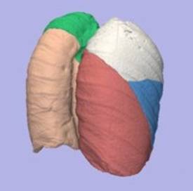
Pulmonary imaging
Lung diseases are one of the main causes of
mortality and disability worldwide. In recent years, hyperpolarized gas MRI has
emerged as a promising new technology with the potential to improve diagnosis
and potentially allow the development of new, better treatments. Before this
promise is fulfilled, advances need to be made in both acquisition technology
and image analysis. We are focusing in the image analysis side by using a
combination of state-of-the-art image analysis methods and computational
models, with the final aim of improving the understanding of image datasets and
developing methods that link image values with the underlying lung function.
Cardiovascular imaging and modelling
|
|
|
Workflow of an automated cardiac histo-anatomy
processing pipeline. In Plank
et al, Phil Trans R Soc A 2009; 367:2257-2292 |
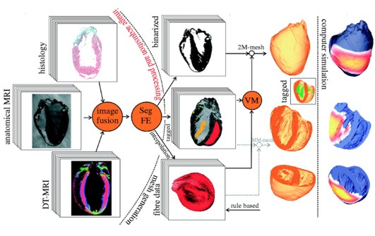
Computational models are becoming a standard tool
in many biomedical applications, and in particular in cardiovascular medicine.
In the last years we have developed a pipeline to build computational models
from high-resolution multimodal images. This includes the development of 3D
histology dataset through registration with MRI scans, segmentation of relevant
structures and mesh generation and the application of ionic models to
investigate the relevance of small structures in electrical simulation results.
Current research includes the extension of these methods to quantify
intersubject variability, through the use of a standardized reference frame.
Biological image processing
Images are becoming ubiquitous in biological
applications. Current image data volumes in biology labs no longer allow traditional
visual analysis, and with the increasing use of high-throughput experiments
there is a pressing need for robust, reusable biological image processing
tools. Our current interests include the use of phase-based operation for
curvilinear structure extraction in microscopy images.