
This page contains links to various resources we create in the course of our work. This includes links to repositories containing data we have collected or helped to collect, software tools that we create implementing our methods, and results that we have produced. The last category includes human brain atlases, tractography recipes, and data sharing collections that contain any aspect of a study that we chose to make openly available.

WIN's Digital Brain Zoo contains MRI data from post-mortem brain samples from a large variety of mammals, including primates, carnivorans, and cetaeceans. The majority of the data are diffusion MRI data, which we use to create white matter atlases. Data were collected by us or our collaborators.
PRIME-DE is an initiative to openly share MRI data, particularly resting state fMRI data, from non-human primates. This project was launched in 2018, following the successful model of the 1000 Functional Connectomes project. Data from Oxford was part of the first release.

Processing MRI data of non-human species means that many standard tools don't quite work. Our MRI Comparative Anatomy Toolbox contains many of the scripts used in the lab to circumvent the problem we encountered. It also contains code to implement some of our analysis methods, including connectivity blueprints (now incorporated into XTRACT), connectivity fingerprint matching, and gradient analyses.

As part of our work comparing different species' brain organization using standardized white matter tracts, we developed a series of tractography recipes describing a large range of white matter tracts in the human and macaque monkey brain (Mars et al., 2018). Based on the original idea of FSL's Autoptx, our library of recipes was joined with a new software wrapper named XTRACT, developed by Saad Jbabdi as part of FSL. This work was done in collaboration with Stam Sotiropoulos and subsequently dramatically expanded upon. Shaun Warrington created the new version of the XTRACT tool (Warrington et al., 2020) and subsequent publications have provided tractography recipes for human infants (Warrington et al., 2022), chimpanzees (Bryant et al., 2020), and gibbons (Bryant et al., 2024), as well as more elaborate recipes for macaques compatible with different templates (Assimopoulos et al., 2024) and reaching subcortical structures.
In a series of papers, we investigated whether it is possible to subdivide parts of the human cerebral cortex based on their connections with the rest of the brain. Here we present the results of these parcellations in the form of atlases for FSLview, part of the popular MRI analysis software package FSL. To install any of the atlases, download the compressed volume below and unpack it into the directory /data/atlases of your FSL version. The atlas should then be accessible in FSLview in the same way as other atlases are. Note that you will be using these atlases strictly at your own risk.
Currently, we have atlases available of lateral parietal cortex (Mars et al., 2011),
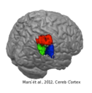
the temporoparietal junction area (TPJ) (Mars et al., 2012),
the dorsal frontal cortex (Sallet et al., 2013),
the ventral frontal cortex (Neubert et al., 2014),
and the cingulate and orbitofrontal cortex (Neubert et al., 2015).
When you use these atlases, please consider referencing the appropriate papers describing the parcellation study.

Some of our publications are associated with code and results shared on the Wellcome Centre for Integrative Neuroimaging's gitlab page. Among others, these include XTRACT protocols for the gibbon (Bryant et al., 2024) and resources from our mouse premotor study (Lazari et al., 2024).

Some of our publications are associated with a Data Sharing Collection on the data repository of the Donders Institute for Brain, Cognition and Behavour. These generally contain analysis code and statistical maps of results.