Software
 |
We have several stand-alone software programs that can help processing data from our and other microscopes.
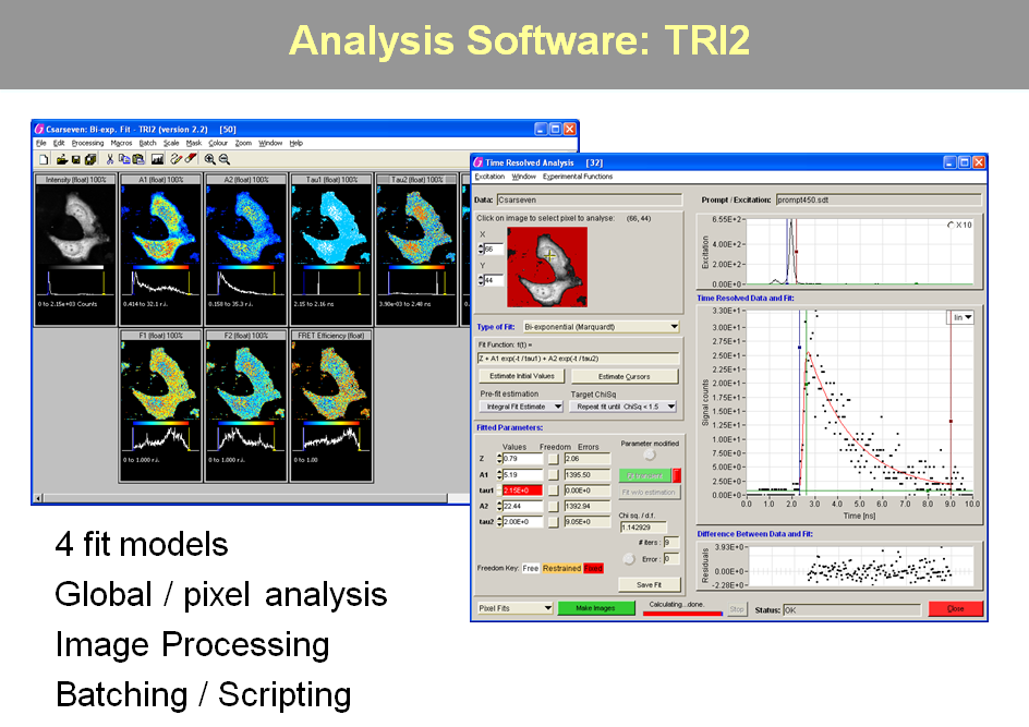
The main purpose of the program TRI2 is to process FLIM images in flexible and novel ways and it has grown to include general image processing functions,
tools for time-resolved anisotropy and methods for the quantitative analysis of histologically stained tissue sections. This works alongside the Mosaic program
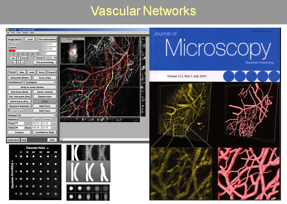
for viewing large images of tissue sections form from tiled images. Trace3D is a program that may help in
delineating and measuring vascular networks.
Some applications are available on the Downloads Page. |
 |
Further Reading
|
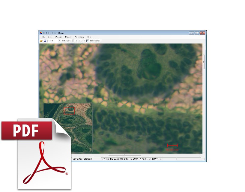
The Mosaic program is an open source application written in C# that can be used to stitch and blend together tiles images to form a seamless mosaic. A windows installer is available from the downloads page and source code from Github |
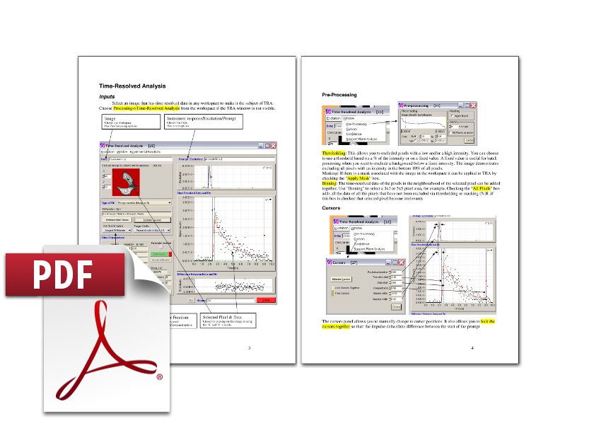
Help document for FLIM processing in TRI2 Non-linear fitting routines are described in this document that are incorporated into TRI2. Further information may be acquired on our wiki and forum for TRI2. |
|
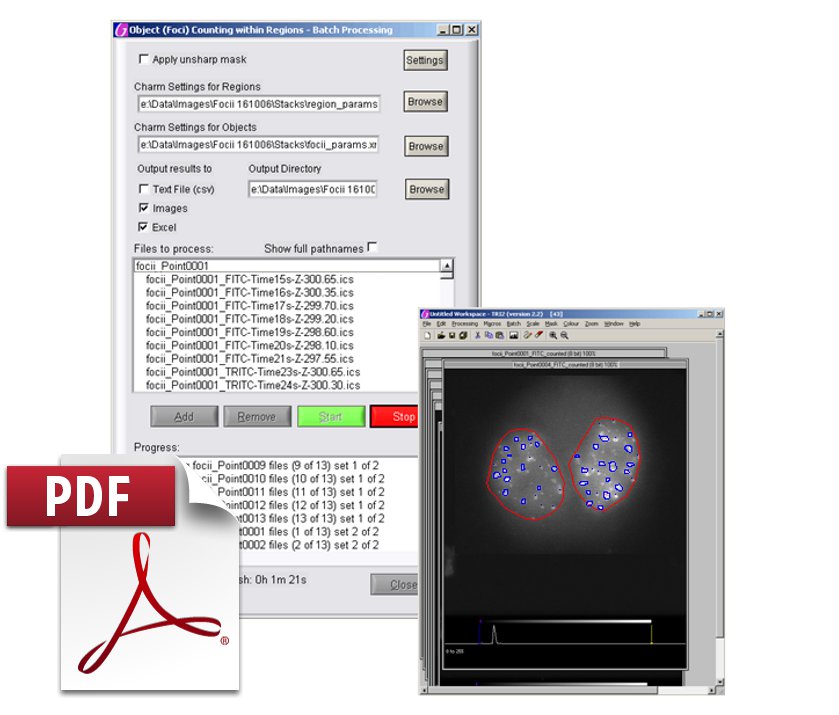
Help document for Batch Counting of foci in TRI2 Details of using TRI2 for foci counting, including some information on how to use the CHARM optimizer to tune the algorithm. [REF] |
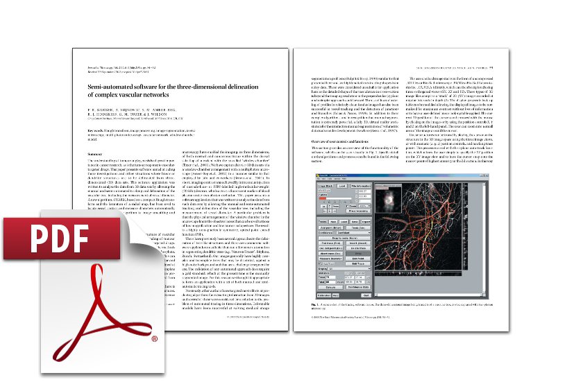
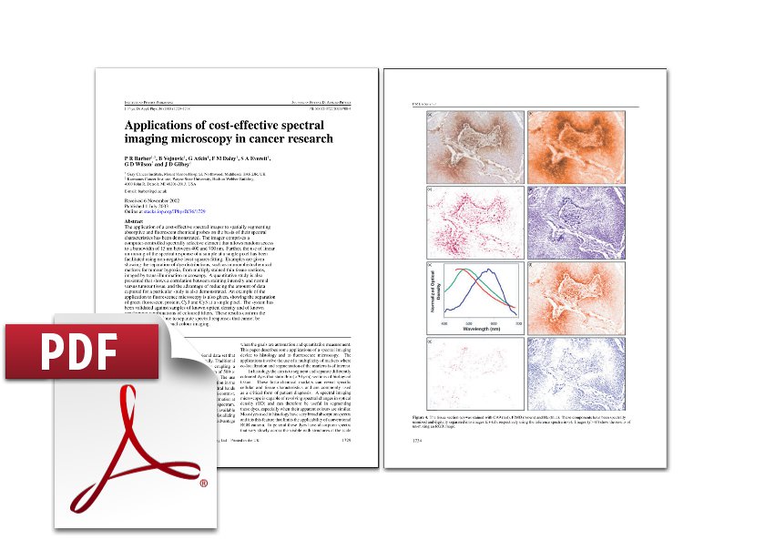
Publication: Barber et al. 2003 Trace3D is a stand-alone program to manually and semi-automatically trace out vascular structures in confocal or 2-photon 3D image stacks. It has then some functions to gather statistics on the structure. |
|
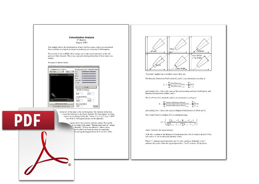
Help document for Colocolisation in TRI2 Here we describe what processing is available for colocalisation analysis in TRI2. |
Publication: Barber et al. 2003 Here we describe what processing is available for separating coloured stains with RGB linear unmixing in TRI2. This is useful for the quantitative analysis of multiply stained histological tissue sections. |
|
Stand-Alone Software Applications You can find out a little bit more about the stand-alone software applications and which may be available for use on Paul's software page. |
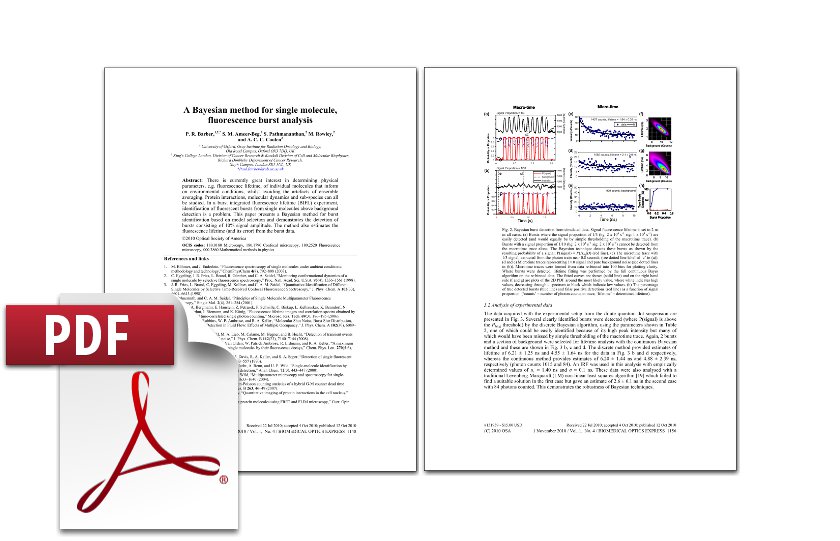
Publication: Barber et al. 2010 Using Bayesian model selection to extract single molecule bursts of fluorescence from noisy data. |