

 Mark Howarth lab
|
|
|
|
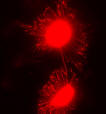
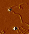
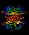
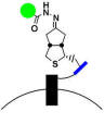
Explanation of figures 1. Part of the Oxford skyline. 2. "Piazza Biochimica" outside our department. 3. Site-specific receptor labeling with quantum dots on carcinoembryonic antigen. 4. The crystal structure of traptavidin, a mutant of streptavidin with 10-fold more stable biotin binding (see papers). 5. An Atomic Force Microscopy image of biotinylated DNA bound to quantum dots, to characterize the number of binding sites (see monovalent QD paper). This image was made together with Andrew Sparks from the lab of Scott Manalis. 6. A way to target small molecules to specific proteins on the cell surface, based upon bio-orthogonal hydrazide conjugation. A collaboration with Irwin Chen (PDF).
|
|
Copyright © 2014 Mark Howarth. All rights reserved. |